
smile-design
Your First Smile Makeover Consultation: What to Expect
Your first smile makeover consultation: what to expect, from the goals discussion to your digital smile design preview and plan.
Sep 20, 20268 min read


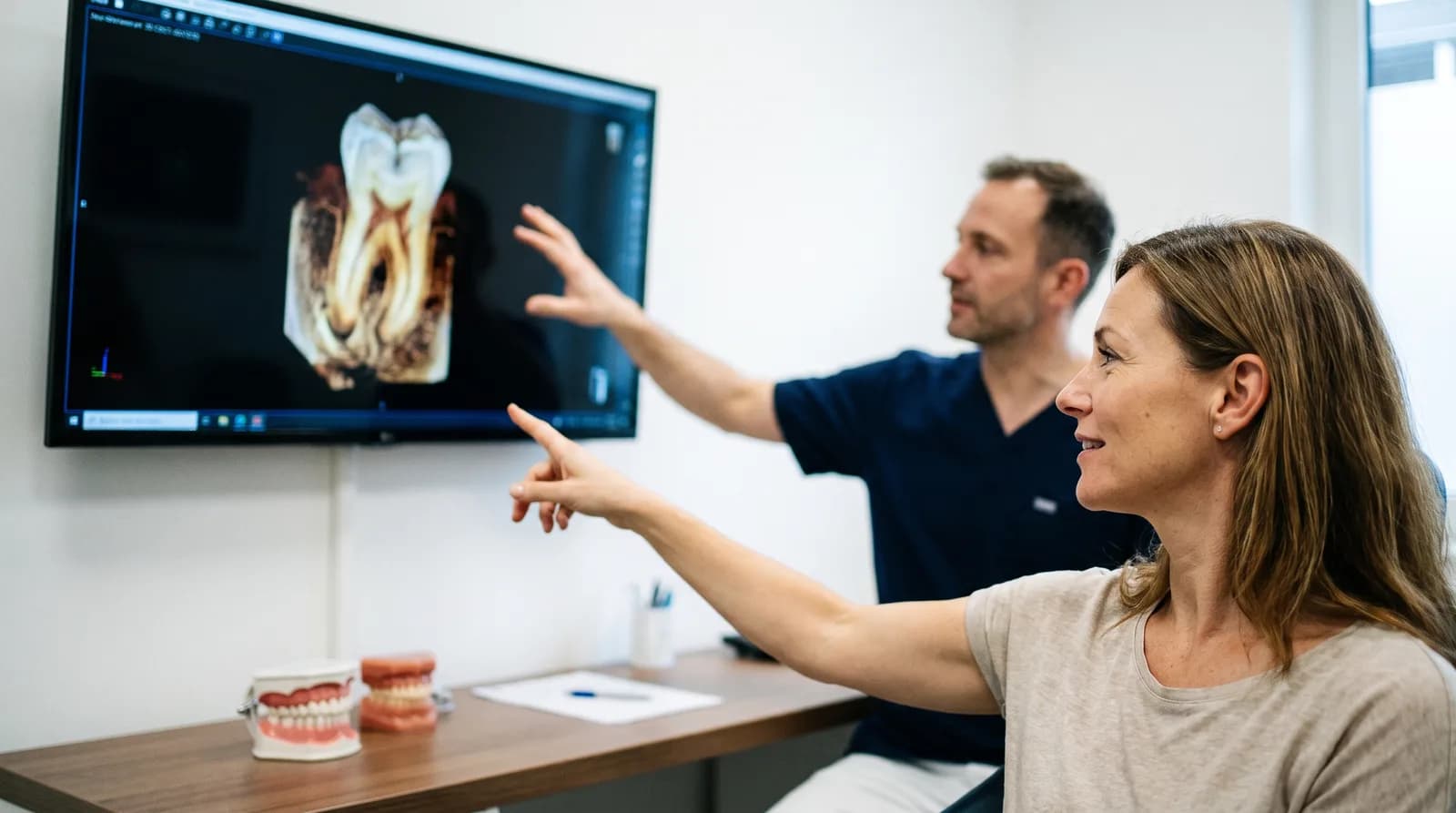
3D endodontics root canal technology uses cone-beam CT imaging to build a three-dimensional map of your tooth's root canal system before treatment ever starts, replacing the flat, two-dimensional x-ray that used to be the only view a dentist had to work with. That shift is a big part of why root canals today look nothing like the procedure most people still picture when they hear the words "root canal."
The reputation these procedures carry is a holdover from decades of limited imaging and less refined instruments, not an accurate reflection of what treatment actually looks like now. Understanding the technology behind modern endodontics goes a long way toward explaining that gap.
Susan J. Curley DDS uses this technology as part of the restorative dentistry care we provide for patients who need root canal treatment. Here's what 3D endodontics actually is, and why it makes a real, measurable difference in how predictable the procedure has become.
3D endodontics refers to using cone-beam computed tomography, or CBCT, to capture a three-dimensional image of a tooth's internal canal system before and during root canal treatment. It replaces the guesswork that came with older two-dimensional x-rays, which flatten a genuinely complex 3D structure into a single angle.
Tooth roots aren't simple straight tubes. Many teeth have curved canals, extra branches, or canals that split and rejoin, none of which show up reliably on a flat x-ray. A CBCT scan captures the full shape from every angle, giving your dentist an accurate map before a single instrument enters the tooth. That map is the foundation everything else about modern endodontics is built on.
Molars are a good example of why this matters. A lower first molar can have anywhere from two to four canals, sometimes with additional branches that a standard x-ray simply won't reveal from a single flat angle. Planning treatment around an incomplete picture used to mean occasionally discovering a hidden canal partway through, or missing one entirely. A 3D scan removes that particular kind of surprise before treatment ever begins, which is a meaningful shift in how the procedure gets approached from the very first appointment.
CBCT imaging makes root canals more precise by showing the exact number, shape, and curvature of canals before treatment begins, which prevents missed canals and reduces the guesswork that led to complications in the past. Seeing the anatomy clearly changes how the entire procedure is planned from the very first step.
A recent systematic review and meta-analysis found CBCT imaging correctly identifies canal morphology with 91% sensitivity and 88% specificity, meaningfully more accurate than relying on two-dimensional imaging alone. Missed canals have historically been one of the more common reasons root canal treatment needed to be redone. Seeing the full anatomy up front, rather than discovering a hidden canal partway through treatment, is exactly what that kind of accuracy prevents.
Root canals earned their reputation decades ago, when dentists worked with limited imaging, hand files, and less predictable anesthesia, which made some procedures longer and less comfortable than they needed to be. Much of that reputation has outlived the technology that caused it, passed down through stories rather than firsthand modern experience.
Older root canal treatment relied heavily on a dentist's tactile sense and a flat x-ray to guess at canal anatomy, sometimes discovering complications mid-procedure that modern imaging would have flagged in advance. Anesthesia techniques have also improved substantially, and rotary instruments move more efficiently through canals than the hand files once used. The procedure most people fear is largely a memory of how root canals used to be done, not an accurate picture of how they're done now.
| Aspect | Older Approach | Modern 3D Approach |
|---|---|---|
| Imaging | Flat 2D x-ray, single angle | 3D CBCT scan, full anatomy |
| Instruments | Rigid stainless steel hand files | Flexible rotary nickel-titanium files |
| Canal length measurement | X-ray estimation | Electronic apex locator |
| Visualization during treatment | Naked eye | Dental operating microscope |
| Missed canal risk | Higher, anatomy discovered mid-treatment | Lower, anatomy mapped in advance |
Today's root canals are more predictable because of CBCT imaging, rotary nickel-titanium files that flex through curved canals more safely than rigid hand files, electronic apex locators that measure canal length precisely, and dental operating microscopes that let the dentist see fine details directly.
None of these existed in their current form when most of the horror stories about root canals were formed. Each piece of technology solves a specific problem. CBCT handles the planning stage by mapping the anatomy in advance. Rotary files move through curved canals with less risk of ledging or breaking than older hand instruments. Apex locators use a small electrical signal to confirm exactly where the canal ends, removing much of the guesswork that used to rely on x-ray estimates alone. Combined, these tools turn what was once an experience-based procedure into a far more measured, verified one.
None of these tools work in isolation. The real shift is how they stack together into a single workflow: imaging informs the plan, rotary instruments execute it efficiently, the apex locator confirms progress in real time, and a microscope lets the dentist verify the result visually before moving to the next step. Each layer catches something the previous one might miss, which is exactly the kind of redundancy that improves outcomes.
Yes, 3D imaging reduces the need for retreatment by catching missed canals, unusual anatomy, and existing complications before the first procedure, rather than discovering them after treatment has already failed. Retreatment is more involved and costly than getting it right the first time.
Procedural errors like underfilled canals, missed canals, or separated instruments have been directly linked to lower success rates in endodontic research. One CBCT-based study found underfilled canals reduce success to around 68%, while separated instruments carry roughly a 14% lower success rate compared to cases without that complication. CBCT imaging addresses this at the planning stage. A dentist can see canal curvature and branching before deciding how to approach the tooth, rather than reacting to a surprise mid-procedure.
Retreatment itself, when it is needed, also benefits from the same imaging. A CBCT scan on a previously treated tooth can reveal exactly why the first attempt didn't fully resolve, whether that's a missed canal, a fracture, or lingering infection the original x-ray couldn't show. That clarity makes retreatment more targeted rather than a repeat of the original guesswork, saving time for both the dentist and the patient.
Curious how this technology applies to your tooth?
Dr. Curley can walk you through what your specific case looks like with modern imaging.
Request an Appointment →Most modern root canals take one to two appointments, each running roughly 60 to 90 minutes, with a single visit becoming more common for straightforward cases thanks to better imaging and instrumentation.
More complex anatomy or existing infection can extend that timeline, but even those cases are more predictable than they used to be. Knowing roughly what to expect going in tends to ease most of the anxiety around scheduling.
Here's what typically affects the appointment count:
Our step-by-step guide to the root canal procedure walks through what happens at each visit in more detail.
Yes, root canal treatment today is generally more comfortable than the procedure most people remember or have heard about, largely due to better anesthesia techniques and more precise, efficient instrumentation. Most patients report the experience feels comparable to getting a filling.
MouthHealthy, the ADA's patient education site, describes root canal treatment as a way to repair and save a tooth rather than remove it, addressing the infection causing the original pain in the first place. Most of the discomfort people associate with root canals actually comes from the infection itself before treatment, not from the procedure that resolves it. Once the infected tissue is removed and the canal is cleaned, the pain that brought someone in usually starts improving right away.
This distinction gets lost in most word-of-mouth accounts of root canals. Someone in significant pain from an infected tooth undergoes treatment, and the relief that follows gets mentally filed under "the procedure was awful" rather than "the infection was awful and the procedure fixed it." Separating those two things out changes the story considerably.
Expect a CBCT scan beforehand to map your canal anatomy, local anesthesia to keep you comfortable, and a procedure guided by that imaging along with rotary instruments and an apex locator to clean and shape the canal precisely. A crown or filling typically follows to protect the tooth afterward.
Here's the general sequence a modern, imaging-guided root canal follows:
Our guide to the signs you need a root canal covers what symptoms typically lead to this point, if you're not yet sure whether that's what you're dealing with. Either way, the version of this procedure your grandparents may have described is largely a thing of the past.
Nervous about a possible root canal?
Modern imaging and instrumentation make today's procedure far more predictable than its reputation suggests.
Book Appointment →3D endodontics root canal technology has quietly changed one of dentistry's most feared procedures into one of its more predictable ones. CBCT imaging, rotary instruments, and precise measurement tools replace guesswork with a clear plan before treatment even begins. If a root canal is somewhere in your future, the version waiting for you looks very different from the one you may be picturing, and the technology behind that difference is exactly why. Results may vary. Please consult with your dentist at Susan J. Curley DDS for personalized treatment recommendations.
Want to see how modern root canal technology works?
Schedule a consultation with Dr. Curley and get a clear picture before you decide anything.
Book Appointment →Think you might need a root canal?
See the signs to watch for →Written By
Dentist
Your first smile makeover consultation: what to expect, from the goals discussion to your digital smile design preview and plan.
What is digital smile design? A preview technology that lets you see your proposed new smile before treatment even begins.
Am I a candidate for veneers? Six clear signs that suggest veneers could be a good fit for your smile and long-term goals.
Book your visit with Dr. Curley online or call us, and we'll take it from there.